Automatic Cell Tracking in Microscopy Images
11 December 2017
Heidelberg researchers develop powerful image analysis method

Source: Karl Rohr / Nathalie Harder (BMCV)
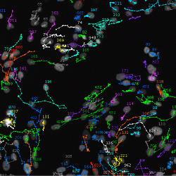
Researchers from Heidelberg University and the German Cancer Research Center have developed a new computer-based image analysis method to automatically track the movement of cells in microscopy images. Using this method, Associate Professor Dr Karl Rohr and Dr Nathalie Harder participated in an international performance comparison in different so-called Cell Tracking Challenges. The Heidelberg cell tracking method yielded the best result in detecting cell cycles used to quantify cell growth and was among the top three with respect to the total number of “top 3 ranks” for different image categories.
The method developed by Dr Rohr and Dr Harder enables quantifying important biological processes such as cell migration and cell growth, both of which play a major role in the development of diseases. The cell tracking method combines segmentation methods with spatio-temporal optimisation to analyse microscopy images. Cells are automatically identified, correspondences determined in consecutive images, and cell divisions detected. The method also determines various information about cell movement such as movement paths, speed, and distance covered.
Karl Rohr heads the Biomedical Computer Vision (BMCV) research group that develops Computer Science methods to automatically analyse cell microscopy as well as radiological images. Dr Rohr's group is located at the BioQuant Center of Ruperto Carola. The group is part of the Bioinformatics and Functional Genomics department at the Institute of Pharmacy and Molecular Biotechnology of Heidelberg University as well as the Theoretical Bioinformatics division of the German Cancer Research Center
The results of the international competition of a total of 21 methods from eleven countries were published in the journal "Nature Methods".

